Digital dental x-rays have come a long way in improving our ability to diagnose problems in your teeth and jaws. But which ones do you need, when do you need them, and why do dentists recommend each kind?
Types of Dental X-rays
Periapical X-rays
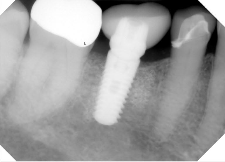
Periapical x-rays capture the full length of 1 or more teeth, so that we can see the apex, or end, of the root. That’s where we look for signs of a tooth abscess, or infection. We also use them, as in this case, to make sure that dental implants are fully integrated in the bone.
Bitewing X-rays
These x-rays of the back teeth are usually taken in groups of 2 or 4, so 1 or 2 per side. Sometimes it may be a group of 7 in a different orientation. Most people have get them every 12 months to check for cavities between the teeth, where we can’t see them. If you’ve never had a cavity between your teeth and are at low risk, your dentist might change it to every 18-, 24-, or rarely every 36 months. Or if you get cavities a lot, your dentist might even recommend them every 6 months. The patient in the below example has gotten a mouthful of dental crowns in her 70+ years. Because we want to catch any cavities around the crowns early, she definitely gets bitewings once a year.

Full-Mouth Series
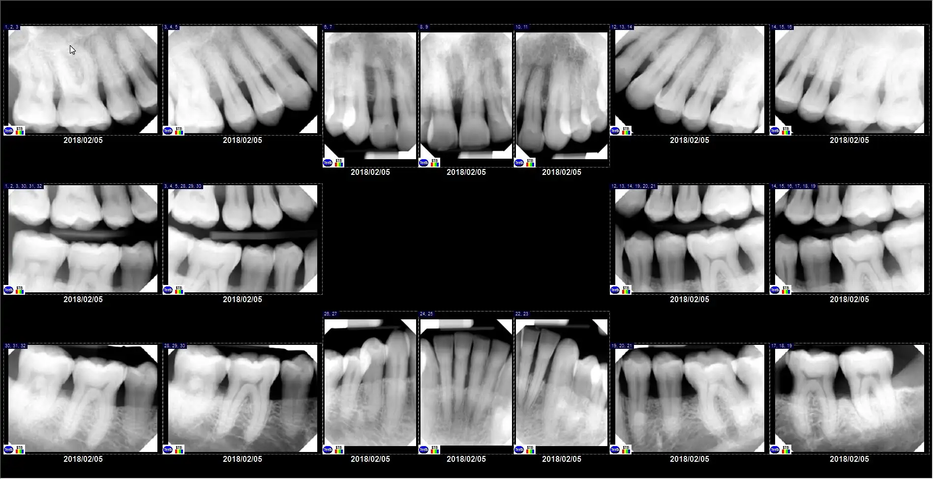
For patients who have cavities on many teeth, or especially if they have gum (periodontal) disease, we take a full-mouth series of x-rays. This is a minimum of 10 total x-rays, including both bitewings and periapical dental x-rays.
Panoramic Dental X-rays
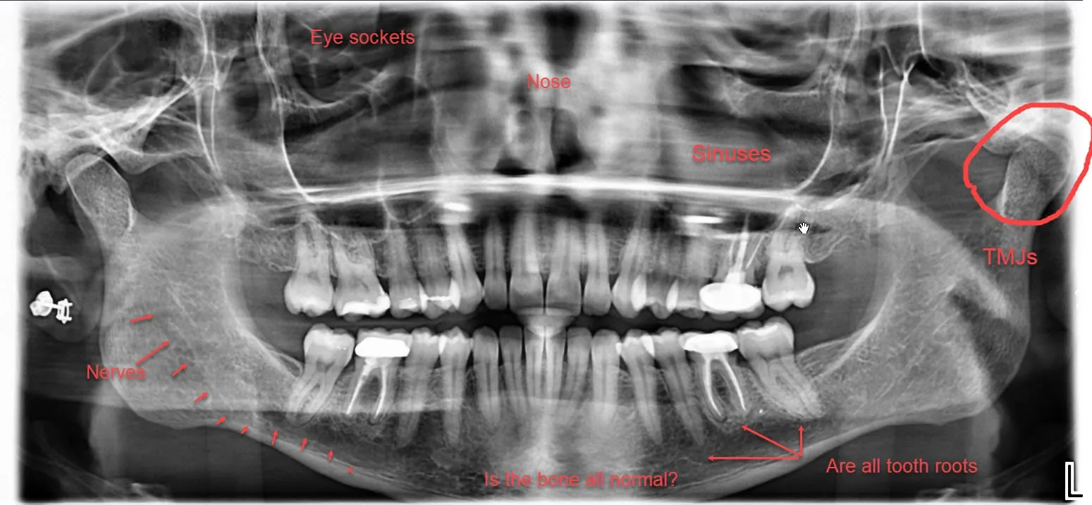
The dental x-rays you see below are called panoramic x-rays. This kind is usually taken ever 3-5 years to screen for possible bone cysts, tumors, sinus abnormalities, etc. It’s also used to check growth and development in children, to make sure their teeth are developing normally. The first one shown here, which I labeled, is of a healthy patient.
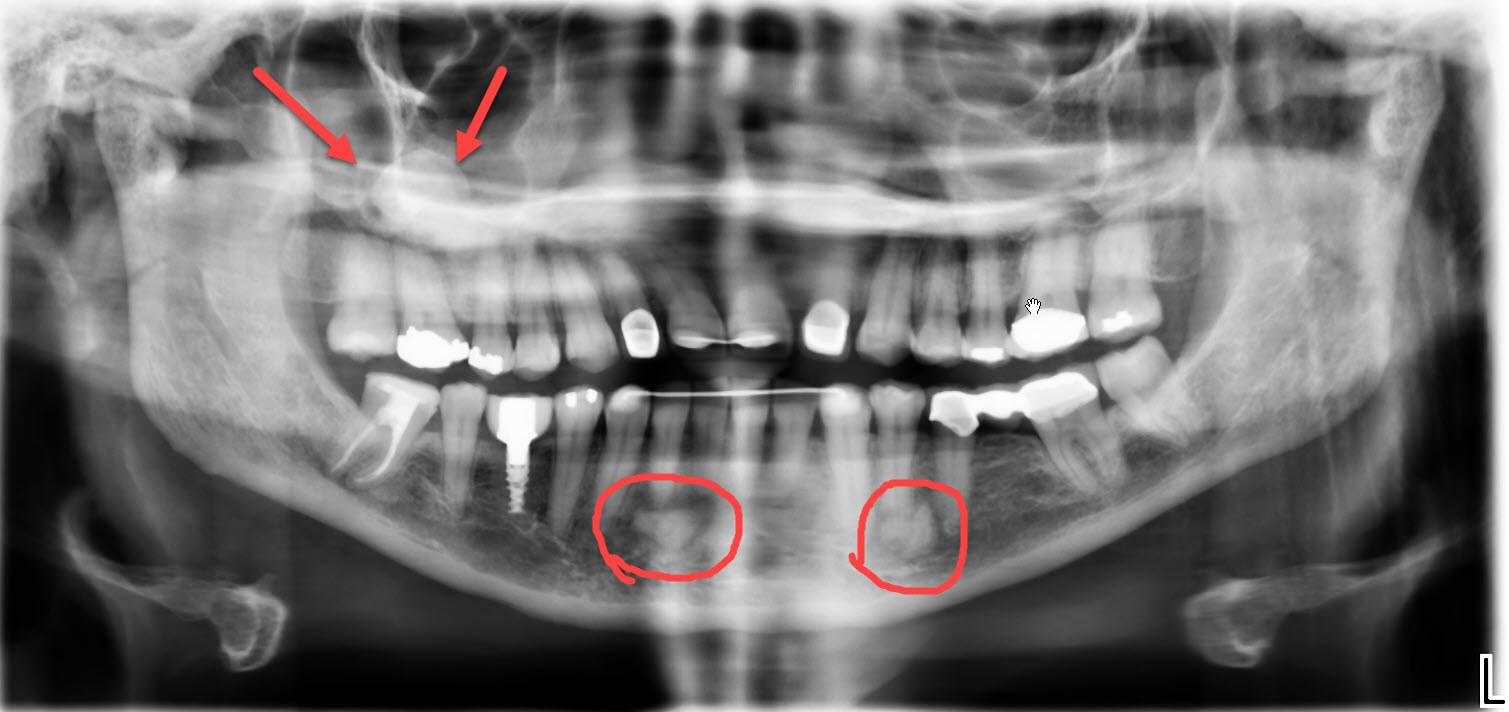
But this panoramic x-ray, taken in the fall of 2019, surprised me with several unusual areas. There’s a large, dense, circular something in the right sinus, which definitely doesn’t belong there. There are also 2 irregular areas of unusually dense bone on the lower jaw. We have sent this patient to an oral surgeon for further evaluation.
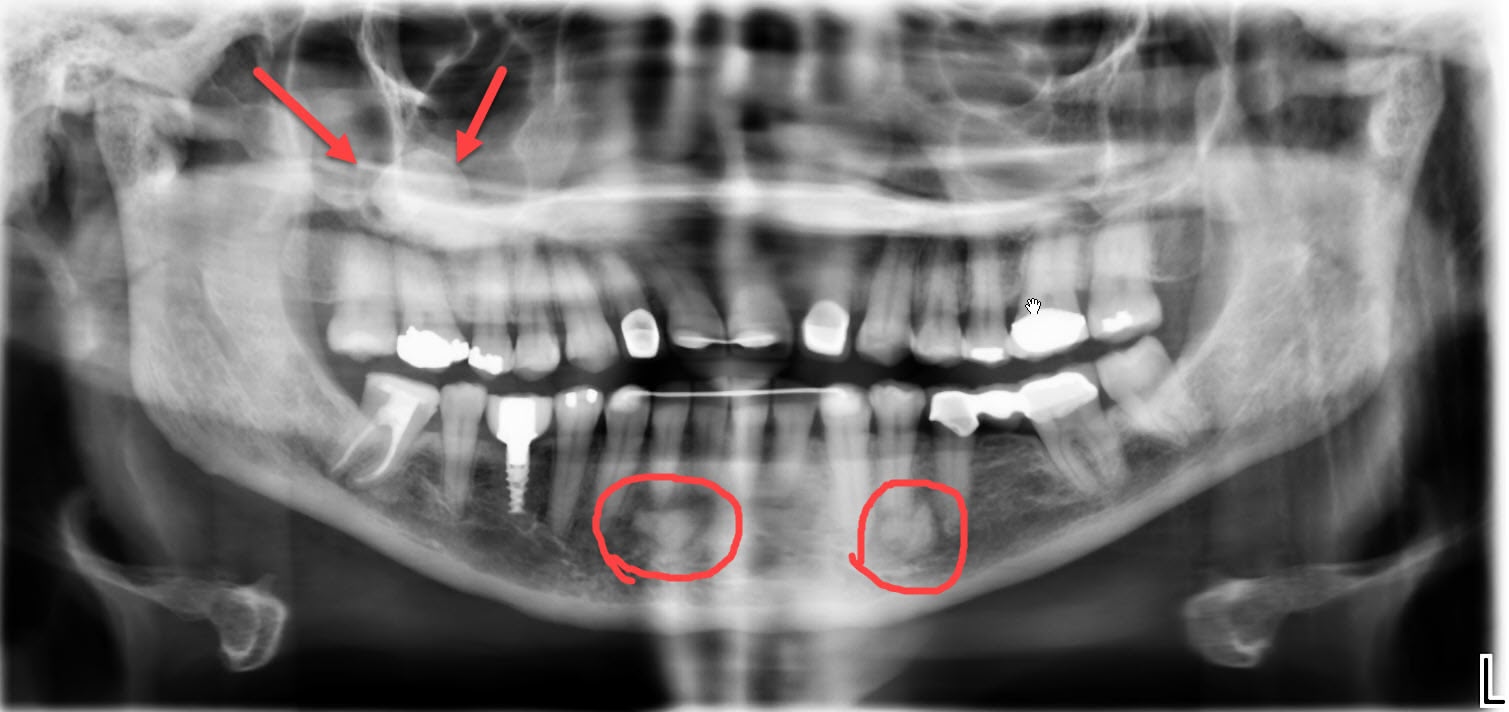
Because we wanted more information about those unusual areas, we took another kind of dental x-ray, described next.
Cone Beam X-ray, or CT Scan
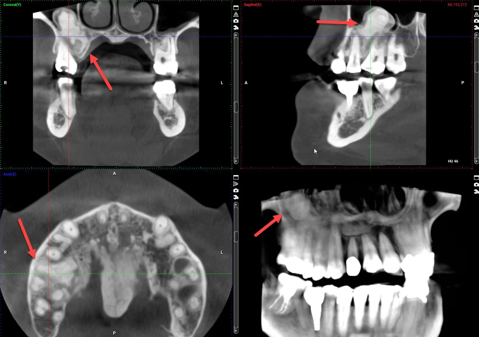
When we need a lot more detail, or different perspectives and angles, we take a CT scan, also called a cone beam or 3D x-ray. Cone beam images are quite different from the other types, because they’re taken in slices. Computer algorithms turn those slices into a 3D images. That 3D x-ray can be rotated in every direction, and we can scan through it up/down, forward/back, and side-to-side. They’re really quite incredible!
These cone beam views are of the same patient above, with the panoramic x-ray. Now we see the true size and shape of those unusual areas.
3D X-Rays for Dental Implants
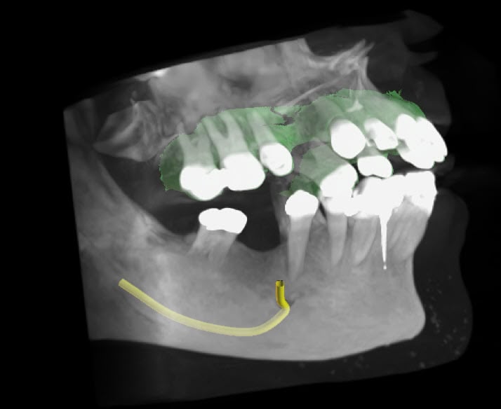
When planning dental implants, we have to know if the bone is both wide and tall enough. If it is, great! If not, then you may need a bone graft or sinus lift to build up the bone. Without the 3D views, we can’t know that. We can also see other important anatomy, like the big nerve in your lower jaw (the yellow line). We really don’t want to run into that!
In short, dental x-rays give us critical information about both your teeth and your jaws. Without them, it’s really impossible for us to do a good job for you.
Call 704-364-7069 or Contact us for a Comprehensive or Emergency Exam.
It’s that easy to start.
