How fast dental cavities grow may sound like a simple question, but it’s actually rather complicated. I decided to write about it, though, after seeing a patient recently to treat a cavity that went from just beginning to an almost-root-canal-sized cavity in just 14 months. It’s a good example of why most dentists still recommend dental bitewing x-rays every 12-18 months.
Is the Cavity There or Not?
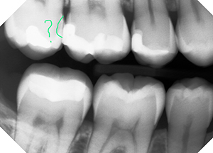
 Here’s the starting x-ray, taken on Dec. 14, 2014. If you look at the circled area, it looks fairly normal. Unfortunately, there was a slight overlap of the teeth in the x-ray, as the digital sensor was angled ever-so-slightly, and the overlap may have masked the area of concern, preventing an earlier diagnosis. Since the patient had few fillings, though, indicated a low risk of cavities overall, we didn’t retake the x-ray to avoid unnecessary exposure. Nevertheless, there’s really nothing there that caused me any concern.
14 Months Later, the Cavity is Obvious & Deep

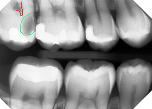
Check out the circled area this time, eh? I’ve outlined the nerve chamber in the tooth on the x-ray in red and the cavity in green, but it’s important to know that a cavity is never sharply defined like this. There are several zones identifiable in cavities, highlighted in the next image. We also know that cavities are typically about 20-30% bigger than can be seen on an x-ray, which is due to 2 reasons:
- The human eye can only distinguish about 60% of the shades of grey as a computer monitor can project, so there is simply detail that the eye can’t detect, and
- The deepest zone of decay has not yet softened enough to be less dense enough for x-rays to detect it, as dental x-rays are all about levels of density. Darker black areas mean it’s extremely soft and x-rays go right through without being blocked, while bright white areas indicate that x-rays are being completely stopped.
Dental Cavities Aren’t Always Obvious from the Outside
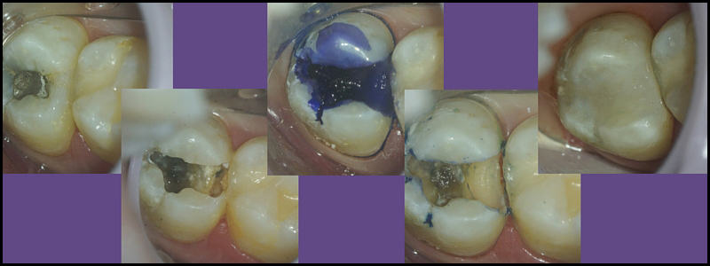
In this short series of photos, taken through our Leica Dental Operating Microscopes at 8.5x magnification, you can see the progression as I clean out the cavity. On the surface, you can’t even tell something is wrong, can you? But once the tooth is opened up, the mushiness is obvious. And when you see how deep the final cavity is, hopefully it also makes sense why dentists tell you that it’s impossible to heal, or reverse, deep cavities, especially with diet.

Not All Dental Cavities Grow Fast. What are the Differences?
As it turns out, there was a good reason that this patient’s cavity grew as fast as it did: the patient had developed a habit of sucking on cough drops due to dry air in his workplace making him cough. He would place the cough drop between his cheek and the upper right molars, and since they weren’t sugar-free cough drops, the long-term sugar exposure in that one area provided the bacteria all the food supply needed to grow rapidly. Obviously, not all cavities will progress at this rate. Sometimes they will progress incredibly slowly, taking years to even break through the enamel and require fixing. If they haven’t broken through the enamel, they might even reverse. Cavities as deep as illustrated, though, can never reverse. They have to be fixed.
Does Everyone Need Dental X-rays Annually?
Most definitely not! While many dentists find it easiest to maintain all their patients on annual bitewing x-rays for consistency (and there’s really nothing wrong with that, as the level of x-ray exposure from dental x-rays is unbelievably small), this is no longer the ADA’s or FDA’s recommendation. In 2012, these organizations developed new guidelines for dental x-rays; while the ultimate decision is still up to the treating doctor, the recommendations allow more flexibility in extending the interval between x-rays, depending on various conditions. The guidelines generally group people into one of three categories, with the corresponding recommended frequencies (these are all for teens and adults; I’ll cover recommendations for kids in an upcoming article):
- New Patients (teens and adults)
- Selected x-rays to visualize areas where decay is most likely to occur.
- This usually includes bitewing x-rays for the back teeth and 2-4 periapical x-rays of the front teeth
- Patients previously or currently diagnosed with periodontal (gum) disease should have a full-mouth series (10-18 x-rays to cover all teeth)
- Selected x-rays to visualize areas where decay is most likely to occur.
- Current Patient (teens & adults) with HIGH Risk or CONTINUING Risk of Dental Cavities
- Selected x-rays every 6-12 months of all areas where decay has recently occurred or is likely to occur
- Current Patient with LOW Risk of Dental Cavities
- Routine bitewing x-rays every 18-36 months
- Quite frankly, the large majority of dentists disagree with 18 months being the routine interval, for the very reason illustrated in this article.
- In our office, we are moving a number of patients to 18-24 month intervals, but every 3 years? Sorry, I’m simply not comfortable with that, because so much can change in 3 years. I don’t actually know of any dentists who do allow up to 3 years between x-rays, although there may be some out there.
To make an appointment for a Complimentary Consultation:
Request an Appointment Online or call us at 704-364-7069.
We’ll look forward to meeting you soon!
- Routine bitewing x-rays every 18-36 months


